
Skip to content
Published surgical research · Aesthetic Plastic Surgery · 2025
Dr. Mert Demirel ↗Instagram ↗
The Chestnut Techniqueby Dr. Mert Demirel
EN

Surgical Technique

The Chestnut Technique

A published breast augmentation approach designed around individualized pocket control, soft-tissue support and implant stability.

Published ResearchResearch led by Dr. Mert DemirelPublished 2025

What is the Chestnut Technique?

The Chestnut Technique is a primary breast augmentation method that combines deliberate implant-pocket sizing with a structured soft-tissue support concept.

In the published description, the implant is supported by a cranial flap superiorly, the pectoralis fascia through the midsection and a caudal flap inferiorly. The fascia and caudal flap are joined to create a defined support architecture around the implant.

The key idea

Chestnut is not a different implant. It is a way of planning the pocket and surrounding support as one integrated system.

Why was it developed?

Breast augmentation is not only a question of implant selection. Pocket dimensions, tissue quality and inframammary-fold control all influence how an implant sits and how the breast may change with time. The Chestnut Technique was described as a structured response to those variables.

What does “stability” mean here?

“Stability” refers to maintaining an intended implant–pocket–fold relationship. It does not mean that other established augmentation techniques are inherently unstable or unsafe. Different approaches can be appropriate depending on anatomy, implant choice and surgeon judgment.

How does the technique work?

The published technique can be understood as a sequence of pocket planning and soft-tissue support steps.

1

Define the dissection boundary

The pectoralis major fascia is dissected to establish the planned superior subfascial boundary.

2

Size the pocket deliberately

The dissection area is checked against the implant base width rather than treated as an open-ended space.

3

Create the support architecture

Cranial coverage, pectoral fascia and a caudal flap are arranged around the implant and joined as part of the construct.

4

Reinforce the fold relationship

The superficial fascia is approximated as an additional fold-support step in the published method.

Published Figure 3. The technique’s cranial, fascial and caudal support architecture.

See the support architecture in motion

The animation translates the published operative concept into a simplified visual sequence, showing how the implant pocket, cranial coverage, pectoral fascia and caudal support relate to one another.

How is it different from established breast augmentation approaches?

Established approaches already use intentional pocket creation, tissue handling and fold control. The distinctive feature of the Chestnut Technique is the way these decisions are described as one defined support architecture. Because the published series did not include a concurrent control group, this comparison describes design emphasis rather than proving superiority.

Clinical questionChestnut TechniqueEstablished approaches
Primary design emphasisPocket sizing and cranial, fascial and inferior support are integrated into one construct.Varies by surgical plane, implant, anatomy and surgeon-specific pocket/fold control.
Inferior supportA caudal flap is intrinsic to the published method.Support strategy depends on plane, tissue quality, fold management and adjunctive techniques.
Pocket sizingThe paper describes checking the pocket against the implant base width.Pocket dimensions are also planned in established surgery, using technique- and surgeon-specific methods.
EvidencePublished 60-patient Level IV clinical series.Evidence varies widely by technique and procedure.

See the operative figures

These four images are the original figures from the publication. Open any figure for a full-screen view, plain-language explanation and the original technical caption.

What does the published study show?

The article describes the operative method and reports a 60-patient clinical series. It documents the technique’s clinical use and the outcomes observed by the authors. It does not establish head-to-head superiority over other augmentation methods.
60patients
2025publication year
Level IVevidence
49:6893–6904journal pages
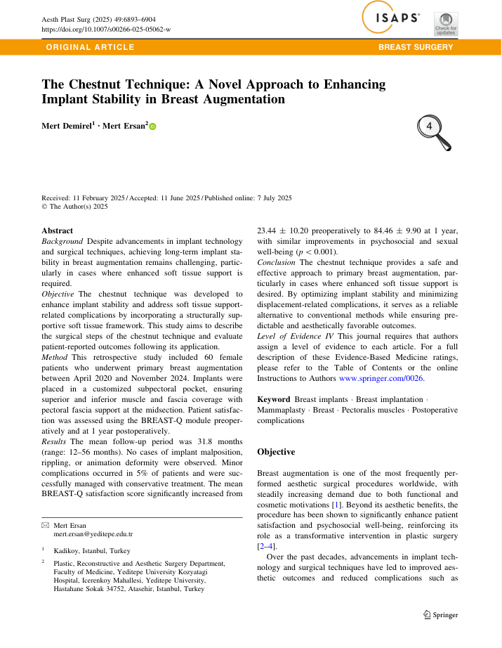
Preview of the published Chestnut Technique paper

The Chestnut Technique: A Novel Approach to Enhancing Implant Stability in Breast Augmentation

Mert Demirel · Mert Ersan
Aesthetic Plastic Surgery · 2025

Life after surgery, in patients’ own moments.

Selected postoperative photographs from patients treated with the Chestnut Technique. The gallery is intended to show real-life patient experiences rather than standardized before-and-after photography.

Individual results vary. Photographs do not predict or guarantee a particular surgical outcome.

What are the limits of the technique and the evidence?

The published paper is best read as a description of a surgical design and an early clinical series — not as a universal rule for every breast augmentation. The study followed 60 selected patients undergoing primary submuscular augmentation, so both the technique's anatomical limits and the study's evidence limits matter when interpreting the results.

01

Implant size and profile are not unlimited

The authors state that the anatomical constraints of the submuscular plane make the technique unsuitable for large-volume implants. They also caution that very high-profile or extreme-profile implants may compromise reliable cranial-flap coverage. In the published cohort, implant volume averaged 335 cc and ranged from 235 to 450 cc; that range describes the patients studied, rather than defining a universal size limit for every person.

02

Breast tissue can still change over time

The technique is designed to support implant position, but it cannot stop ageing, pregnancy, breastfeeding, weight fluctuation or changes in tissue quality. The paper specifically discusses the possibility of breast tissue migrating downward over a relatively stable implant — sometimes described as waterfall or snoopy deformity. This was not observed in the published cohort during follow-up, but the authors note that it can remain a longer-term possibility.

03

Bleeding and fluid collections remain possible

Creating and elevating the caudal flap introduces a region in which bleeding, hematoma or seroma can occur if hemostasis is not meticulous. Three of the 60 patients in the series developed an early localized hematoma or seroma; all were managed with local or outpatient drainage and did not require further surgery. The paper therefore treats careful vessel control as an important technical part of the operation, not as a risk that the technique eliminates.

04

The study is descriptive, not a head-to-head trial

The evidence is Level IV and comes from a retrospective case series without a concurrent control group. That means the study can report what happened in this cohort, but it cannot prove that the Chestnut Technique has lower complication rates or is superior to dual-plane, subfascial or other established approaches. The authors explicitly call for larger studies, longer follow-up and multi-surgeon evaluation.

05

The published population was deliberately selected

The study focused on primary submuscular augmentation. Revision cases, patients planned for subglandular or subfascial augmentation, patients with a history of breast disease, and those without at least one year of follow-up were excluded. Two implant brands were used, but the study was not powered to compare them. The published findings therefore apply most directly to a defined primary-augmentation population rather than every possible breast-implant scenario.

Ten questions about the Chestnut Technique

These answers translate the published paper into patient-readable language. They explain the design logic and the results reported by the authors, while keeping the difference between a published clinical series and an individual surgical recommendation clear.

Is the Chestnut Technique a special type of breast implant?

No. The Chestnut Technique is a surgical method for designing the implant pocket and using the patient’s own muscle and fascial tissues as a structured support system. The published series included both Mentor and Motiva implants, so the concept is not tied to one proprietary implant. The paper did not compare brands against each other; its emphasis is on how the implant is supported and positioned rather than on a specific manufacturer.

Why is it called the “Chestnut” Technique?

The name comes from the surgical anatomy described in the paper. Once the implant is positioned, the superior and inferior tissue flaps partly surround it in a way the authors compare to a chestnut emerging from its shell. The name is therefore not a marketing reference to shape; it is an analogy for the relationship between the implant and the cranial and caudal support layers.

What does “implant stability” mean — and does it mean other techniques are unstable?

Stability refers to maintaining the intended implant position and the integrity of the lower breast fold over time as tissue is exposed to implant weight, friction, gravity and movement. Problems such as bottoming-out, displacement, double-bubble deformity, rippling or asymmetry can occur after breast augmentation for many reasons. Established techniques can be safe and effective; the Chestnut paper proposes one specific structural strategy for this problem and, because there was no control group, does not prove that other approaches are unsafe or inferior.

What is the most distinctive part of the technique?

The central idea is a three-part support architecture: cranial coverage above the implant, pectoral fascia across the midsection and a caudal flap below. The caudal flap is intended to provide relatively static support, while the cranial flap contributes more dynamic coverage. The pectoral fascia is then sutured to the caudal flap, creating continuity across the pocket rather than relying on a single tissue plane alone.

Why does pocket size and positioning matter so much?

In the published method, the pocket is deliberately defined around the implant rather than being broadly dissected and then accepting whatever space remains. A paper ruler cut to the implant’s base width is used intraoperatively to help define the superior and inferior boundaries, while important medial and lateral attachments are preserved. The broader principle is that implant selection and pocket design should follow the patient’s anatomy, breast footprint and fold position instead of forcing anatomy to fit a predetermined implant.

Can the Chestnut Technique be used with very large or very high-profile implants?

Not necessarily. The authors explicitly describe large implant volume as an inherent limitation because the submuscular anatomy restricts the available space for the pocket. They also caution against extreme profiles when the cranial flap may not maintain adequate coverage. The published series used implants from 235 to 450 cc, with a mean of approximately 335 cc, but those numbers should be understood as the study range rather than a universal prescription or cutoff.

Is every breast-augmentation patient a candidate?

No. The paper describes selected patients undergoing primary submuscular breast augmentation, not every clinical scenario. Revision operations, planned subglandular or subfascial cases, patients with a history of breast disease and several other groups were excluded from the study. In practice, breast-tissue thickness, inframammary-fold anatomy, chest-wall asymmetry, implant goals, previous surgery, medical history and the findings of an examination all influence whether this technique is appropriate.

What results were reported in the published series?

The paper reports 60 patients with a mean follow-up of 31.8 months, ranging from 12 to 56 months. During that follow-up, the authors observed no implant malposition, animation deformity or rippling, and no clinically significant implant-palpability problems were reported. Three patients — 5% of the cohort — developed an early localized hematoma or seroma that was treated with drainage. These are the outcomes of this particular case series; they are not a guarantee of the same result for every future patient.

How was patient satisfaction measured?

The study used the validated BREAST-Q Augmentation Module before surgery and again at one year. Mean “satisfaction with breasts” scores increased from 23.44 before surgery to 84.46 at one year; psychosocial and sexual well-being scores also improved significantly, while physical well-being remained statistically stable. All 60 patients completed the one-year assessment, and the paper states that an independent physician who was not involved in the operations performed the evaluations.

Where can I see the technical details, and how do I know whether the technique is relevant to me?

The four published journal figures and the surgical animation are available on this site for readers who want to go deeper into the operative design. The publication is cited here by title, authors, journal and year, but this site intentionally does not provide a PDF, DOI link or publisher access link. These materials explain the technique, but individual suitability still requires assessment of anatomy, tissue quality, implant goals, health history and alternative surgical options during a clinical consultation.

Dr. Mert Demirel at a professional meeting

Research led by Dr. Mert Demirel

The Chestnut Technique is directly connected to Dr. Mert Demirel’s clinical and academic work in aesthetic breast surgery. This site is a focused patient-and-research guide to the published technique.

Visit Dr. Mert Demirel’s main website ↗

Research information is not an individual recommendation.

A published technique can explain a surgical concept, but suitability still depends on anatomy, tissue quality, implant goals, medical history and clinical examination.

Could the Chestnut Technique be relevant to your anatomy?

The answer depends on individual anatomy, tissue support, desired implant dimensions and surgical goals.

Review the study evidence ↑
4.8 Google · 132 reviews Want to learn more about the Chestnut Technique?Ask the clinic about the technique.